Moby Parsons, MD
3D imaging technology has led to a much better understanding of glenoid morphology and how it is affected by the wear process in shoulder arthritis. The pathologic triad as described by Matsen1 (1. posterior humeral subluxation; 2. increased glenoid retroversion; 3. biconcave glenoid) is encountered in many arthritic shoulders. Other wear patterns like superior erosion may also commonly occur in certain conditions like cuff tear arthropathy. One of the principle goals of shoulder replacement, whether anatomic or reverse, is to recognize and correct pathologic glenoid deformity as failure to do so may risk premature loosening of the glenoid implant due to abnormal loading mechanics.
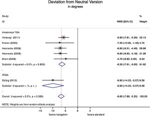
Unfortunately, even experienced shoulder surgeons do a poor job in correcting glenoid erosion. A meta-analysis by Sadoghi et al demonstrated an average error in glenoid correction of +/- 11 degrees2. Other research by Iannotti et al showed an angular variability of 10 degrees in pin placement using a free-hand technique3.
Advanced CT imaging has allowed surgeons to preoperatively plan the placement of the glenoid component with the goal of correcting pathologic version, minimizing bone loss and preventing penetration of the glenoid vault.
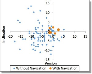
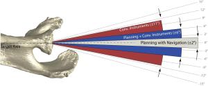
This lack of precision is no longer acceptable given today’s technology. Advanced CT imaging has allowed surgeons to preoperatively plan the placement of the glenoid component with the goal of correcting the pathologic version, minimizing bone loss and preventing penetration of the glenoid vault. As many systems now offer augmented glenoid implants, such systems also allow selection of the optimal implant for each given case. Research looking at the ability of surgeons to recreate a preoperative plan using conventional, free-hand instruments compared to surgical navigation has been performed. The results demonstrate that even with planning, a surgeon’s ability to execute that plan remains very inaccurate. The scatter plot above shows the range of implantation variability without navigation in blue compared to with navigation in orange. These results clearly show that eye-balling it in the operating room is no longer acceptable with today’s technology.
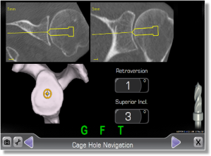
The ExactechGPS platform is a surgical navigation system that allows the surgeon to register the patient’s anatomy with their CT scan. Trackers attached to the scapula and the instruments then allow the surgeon real-time visualization of glenoid preparation so that the preoperative plan can be replicated with great precision. Navigation can help surgeons place and orient the central cage hole, the reaming of the glenoid surface and the placement of fixation screws in the reverse shoulder.
Particularly in complex cases with severe retroversion, severe erosion or a very narrow glenoid vault, navigation can improve the accuracy and precision of glenoid component placement. Some cases have very little margin for error and given the demands that today’s patients place on their implants, it is up to surgeons to maximize outcomes and durability using modern navigation technology.
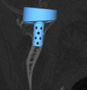
The picture below shows improved precision that ExactechGPS can provide the shoulder arthroplasty surgeon4. This is the only system that currently allows real-time navigation of a preoperative plan which differentiates it from patient-specific instrumentation.
Moby Parsons, MD, is in private practice in New Hampshire. He completed his internship and residency at University of Pittsburgh and fellowships at the University of Washington, Southern California Orthopaedic Institute and the University of Sydney. His work has produced multiple published articles and international presentations.
References:
1. Matsen, F. A. et al. (2014). Clinical orthopaedics and related research, 473(6), 2088-96.
2. Sadoghi, P. et al. Arch Orthop Trauma Surg (2015) 135:41–47
3. Hendel, M. et al. The Journal of Bone & Joint Surgery. 94(23):2167–2175, Dec 2012
4. Greene, A. et al. Navigated vs. non-navigated results of a CT based computer assisted shoulder arthroplasty system in 30 cadavers. Presented at ISTA 2018