Kaveh R. Sajadi, MD
Read complete study: Bone Graft Augmentation for Severe Glenoid Bone Loss in Primary Reverse Total Shoulder Arthroplasty
Following the legacy of hip and knee replacements in relieving pain and improving function for patients with arthritis, shoulder replacements are now the fastest growing joint replacement. Shoulder replacements reliably improve the quality of life for most patients and approximately 85% are still in place 15 years later. The leading cause of failure is loosening of the component on the glenoid side of the shoulder. Minimizing this improves longevity and outcomes.
Shoulder arthritis is often characterized by significant glenoid bone loss. Classically, primary arthritis is associated with posterior glenoid wear, while rotator cuff tear arthropathy leads to superior glenoid wear. To minimize the risk of glenoid loosening, it is important to restore glenoid alignment and version and obtain secure fixation. The surgeon may be faced with managing diminished bone stock or significant deformity to achieve these goals. The primary options for managing bone loss on the glenoid side are eccentric reaming, bone grafting, and augmented glenoid implants.
In “Bone Graft Augmentation for Severe Glenoid Bone Loss in Primary Reverse Total Shoulder Arthroplasty,” Dr. Lorenzetti and his coauthors have reported their outcomes with one of these approaches, bone grafting, in conjunction with reverse shoulder arthroplasty. The authors had two goals: first, to determine the outcome of reverse arthroplasty when bone grafting was performed as part of a primary procedure; second, to correlate outcomes with the amount of native bone support at implantation. They conducted a retrospective of 57 patients with minimum 2-year follow up who had this procedure done.
In addition, they used CT scans preoperatively and plain radiographs postoperatively to measure the degree of bone loss and the amount of native bone support under the glenoid baseplate. Replacements were performed for rotator cuff tear arthropathy, rheumatoid arthritis, osteoarthritis, and chronic dislocations. Due to the diversity of diagnoses, the wear pattern varied. ASES, Simple Shoulder Test, VAS pain and function scores, and patient satisfaction were assessed at minimum 2-year follow up.
Intraoperative decision making determined that if at least 80% of contact could not be achieved between the baseplate and native glenoid, bone grafting was utilized. When available and of sufficient quality, humeral head autograft was preferentially used. Otherwise, femoral head allograft was chosen. Virtual models of the bone and implant were created based on preoperative CT and postoperative x-rays and used for virtual implantation to determine the baseplate contact with native bone, which was reported as a percentage of total surface area available. A previously performed cadaveric study was used as a validation for this virtual technique.
The authors reported significant improvements in ASES scores, patient satisfaction, simple shoulder test, range of motion, and pain scores. Further, they documented graft incorporation in 98% of patients. The mean contact area percentage of the implant on native bone was 17%. They did not find an association of outcome with bone contact area.
I commend the authors on their technique and outcomes. Their high incorporation rate and low failure rate show that bone grafting for significant glenoid defects is a successful technique. It is important to consider some factors not specifically mentioned though.
First, restoration of normal glenoid alignment is an important goal of reverse arthroplasty, and the authors do not report pre- or postoperative version. Second, graft preparation and sizing should optimize this restoration and shaping the glenoid bone graft can be challenging. Finally, although contact with native bone is measured, the amount of fixation of the implant into native bone is not described. The implant must make significant purchase, generally a centimeter or more, in the native glenoid, often through the graft, to achieve stable fixation. Having a long central post/cage or screw facilitates this.
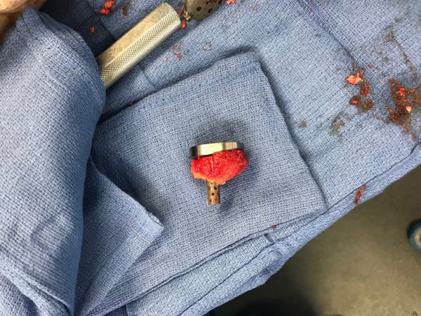
I have performed this technique with success as well. The pictures below are of a 71-year-old gentleman with a Walch B2 glenoid with approximately 25 ° of posterior glenoid wear. I chose to use humeral head autograft with a long central post implant to insure fixation in native bone. Preoperative and postoperative imaging are included.


While this paper describes one particular method, it is important to remember there are other ways to manage glenoid bone loss.
Eccentric reaming is a simple technique that can be used to manage smaller amounts of glenoid retroversion or superior wear, up to 10-15 °. Another method utilizes augmented glenoid implants to manage the bone wear. Augmented implants are now available for both anatomic and reverse arthroplasties, and are available in posterior, superior, and posterosuperior options. Prosthetic augments can also be combined with bone grafting to better restore normal version and alignment. Augments in isolation remove the concern for grafting, including incorporation or resorption or the risk of infection with allografts. Finally, guidance systems are available to help with implantation in appropriate alignment in cases with significant bone loss. Some systems provide 3D printed models to be used intraoperatively, while another provides live intraoperative computer-assisted navigation to insure proper implant and screw positioning.
When taking on a complex case with glenoid bone loss, careful preoperative planning is essential. This includes review of imaging (including CT scan), having graft options available, and choosing the right implant system for the case – one that has the right options for the problem at hand.
Kaveh Sajadi, MD, practices orthopaedics with Kentucky Bone and Joint Surgeons and is an instructor in the University of Kentucky’s residency program. He completed his residency at the Campbell Clinic and his fellowship at the NYU Langone Orthopedic Hospital. Dr. Sajadi is a frequent instructor at Exactech domestic and international shoulder courses.